What Split-Brain Patients Reveal About Consciousness
The brain, like the rest of the body, has a remarkable degree of bilateral symmetry. It is helpful to think of it as an enlarged walnut. One side is not the exact mirror image of the other, but approximately so. Almost every brain structure has two copies, one on the left and one on the right. The left side of the visual field is represented by the visual cortex in the right hemisphere, whereas the right side is mapped onto the left visual cortex.
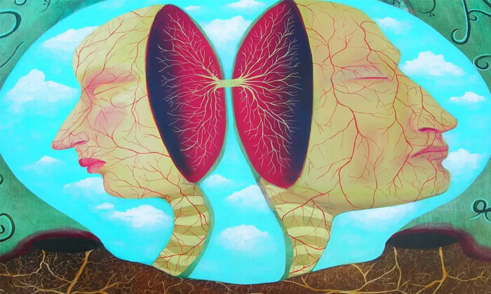
Yet when you look out at the world, you don’t see a fine vertical line running down your field of view; the two hemifields are integrated seamlessly. Philosophers emphasize that experience is unitary. That is, you don’t experience two streams of consciousness, one for each side, but only one. And what is true for vision applies with equal force to touch, to hearing, and so on.

When I mention Descartes’ identification of the pineal gland in class, some students snicker, “How silly.” In fact, Descartes was centuries ahead of his time in his search for a relationship between structure and function. He’s a breath of fresh air — of modernity and enlightenment — in the dusty, moth-eaten atmosphere of the closing years of Medieval scholasticism. Descartes replaced worn-out Aristotelian teleonomic, final causes that really don’t explain anything — for example, wood burns because it possesses an inherent form that seeks to burn — with mechanistic ones.
The discordance between the two halves of the brain and the one mind was first pointed out by René Descartes, who, in the 17th century, sought a single organ that would reflect the unitary nature of experience. He erroneously assumed that the pineal gland has no left and right halves and famously hypothesized that it was the seat of the soul (or, in modern terms, the neural correlate of consciousness).
But for all of Descartes’s insights, he failed to recognize that it is the corpus callosum, the largest white matter structure in the brain, that is primarily responsible for the integration between its two hemispheres. It is a thick bundle, a bit like a ribbon cable, of about 200 million axons, each extending from a pyramidal cell on one side of the brain to the other side. These axons, together with some minor wire bundles, tightly coordinate the activities of the two cerebral hemispheres so that they work together effortlessly, giving rise to a single view of the world.
What happens if this bundle of axons is cut? If it were done carefully, without damaging other structures, the patient should remain sentient, though his or her consciousness might be split in two, shrinking to encompass only the left or right visual field, with the other half invisible. However, this is not what happens!
Consider cases of intractable epileptic seizures, when the entire corpus callosum is cut to prevent a seizure that originates in one hemisphere from spreading into the other and causing generalized convulsions. This operation, first performed in the early 1940s, alleviates seizures.
One half of the brain quite literally does not know what the other half is doing.
Remarkably, once these “split-brain” patients recover from the surgery, they are inconspicuous in everyday life. They see, hear, and smell as before; they move about, talk, and interact appropriately with others, and their IQ remains unchanged. They have their usual sense of self and report no obvious alteration in their perception of the world — no shrinkage of their visual field, for example. The surgeons who pioneered this operation, including Joseph Bogen at Loma Linda University in Southern California, were puzzled by this lack of clear symptoms.
Closer inspection of split-brain patients by the biologist Roger Sperry at Caltech, however, revealed a persistent and profound disconnection syndrome. If specific data are given to one hemisphere, that information is not shared with its twin on the other side. Furthermore, only one hemisphere, typically the left one, speaks. That is, if the right hemisphere is lost or silenced by anesthesia, the patient can still talk, which is why the left hemisphere is called the dominant hemisphere. The right hemisphere by itself has only limited language comprehension and is mute, though it can grunt and sing. So, when engaged in conversation with a split-brain patient, it is the person’s left hemisphere that is doing all the talking. The patient can’t name an object presented in the left visual field because that image is processed by his mute right visual cortex. But they can pick out an object from a group on a tray using their left hand, which is controlled by the right motor cortex.
If a key is placed in the patient’s right hand, with the hand under the table and out of sight, they will quickly name it. Touch information from their right hand is transmitted to their left hemisphere, where the object is identified, and its label is relayed to the language center. If the key is placed in the person’s left hand, however, the patient is unable to identify it and rambles on. The right hemisphere might very well know that the object is a key, but it cannot convey this knowledge to the language centers on the left, because the communication links have been cut.
In other words, one half of the brain quite literally does not know what the other half is doing, which leads to situations somewhere between tragedy and farce.

Victor Mark, a neurologist at the University of North Dakota, videotaped an interview with a split-brain patient. When asked how many seizures she had following her operation, her right hand held up two fingers. Her left hand then reached over and forced the fingers on her right hand down. After trying several times to tally her seizures, she paused, then simultaneously raised three fingers with her right hand and one with her left. When Mark pointed out this discrepancy, the patient commented that her left hand frequently did things on its own. A fight ensued between the two, looking like slapstick comedy. Only when the patient grew so frustrated that she burst into tears was I reminded of her sad situation.
Studies with split-brain patients, work for which Sperry was awarded the Nobel Prize in 1981, teach us that cutting the corpus callosum cleaves the cortico-thalamic complex in two but leaves consciousness intact. Both hemispheres are independently capable of conscious experience, one being much more verbal than the other. Whatever the neural correlates of consciousness are, they must exist independently in both hemispheres of the cerebral cortex. Two conscious minds in one skull. From the point of view of one of these minds, the other one is inaccessible; it may as well be on the dark side of the moon (although the two hemispheres can cue each other unconsciously).
As long as you are awake, you are conscious of something — the road ahead, a heavy metal piece by Rammstein running incessantly through your mind, or fantasies about sex. It is only during certain meditative practices that one can be conscious without having any specific content, conscious without being conscious of anything (pure or naked awareness). Even when your body is asleep, you can have vivid experiences in your dreams. In contrast, during deep sleep, anesthesia, fainting, concussion, and coma, there is no experience at all. Not a black screen, but nada.
When severe injury strikes the brain, for example, consciousness may not return. A car accident, a fall, a combat wound, a drug or alcohol overdose, a near drowning — any of these can lead to seemingly profound unconsciousness. Thanks to rescue helicopters and emergency medical technicians, who quickly deliver the victim to the care of a team of specialized trauma nurses and physicians with their advanced tools and pharmaceutical cornucopia, many can be plucked back from the edge of death. Although this is a blessing for most, it is a curse for a few. Some remain alive for years yet never recover the ability to speak or otherwise interact with their family or medical personnel.
These global disorders of consciousness occur when the brain regions responsible for arousal are damaged. Neurons in the thalamus and cerebral cortex can’t assemble into the far-flung neuronal coalitions that mediate any one conscious content. Impaired states of consciousness include coma, the vegetative state, and the minimally conscious state. Overall arousal fluctuates from complete absence in coma, to periodic sleep–wake transitions in the vegetative state, and awakenings with purposeful movements in the minimally conscious state, sleepwalking, and certain partial epileptic seizures.
During deep sleep, anesthesia, fainting, concussion, and coma, there is no experience at all. Not a black screen, but nada.
In the United States alone, as many as 25,000 patients hover for years in a vegetative state termed persistent vegetative state (PVS), with bleak prospects for recovery. What makes the situation almost unbearable is that, unlike comatose patients, who exhibit almost no reflexes, patients in this limbo state have daily sleep–wake cycles. When they are “awake,” their eyes are open and may move reflexively, as do their limbs on occasion; they may grimace, turn their head, or groan. To the naïve bedside observer, these movements and sounds suggest that the patient is awake, desperately trying to communicate with their loved ones. The tragedy of the ruined patient’s blank and empty life, drawn out over hopeless decades in hospices and nursing homes, is mirrored and amplified by the love — and the resources — their family expends on her care, always hoping for a miraculous recovery.
You may recall Terri Schiavo in Florida, who lingered for 15 years in a persistent vegetative state until her medically induced death in 2005. Because of the nasty, public fight between her husband, who advocated discontinuing life support, and her parents, who believed that their daughter had some measure of awareness, the case caused a huge uproar, was litigated up and down the judicial chain, and eventually drew in then-President George W. Bush. Medically, her case was uncontroversial. She had brief episodes of automatisms: head turning, eye movements, and the like, but no reproducible or consistent, purposeful behavior. Her EEG was flat, indicating that her cerebral cortex had shut down. Her condition failed to improve over many years. The autopsy showed that her cortex had shrunk by half, with her visual centers atrophied; so, contrary to public reports circulating at the time, she couldn’t have seen anything.
In acute patients in the intensive care unit (ICU) that are deemed to be unconscious based on behavioral measures (that is, asking the person to speak or move their limb or eyes or pinching their fingernail to see whether they withdraw the hand in response to this painful stimuli), the situation is more critical since if they remain unresponsive the clinical team will, within a few days, initiate discussion with the family regarding whether the patient would have wanted to continue to live under these dire conditions. For most of these, the loved ones agree that life support should be withdrawn, and the patient dies.
Unfortunately, distinguishing between a patient in a vegetative or behaviorally unresponsive state, who has regular sleep–wake transitions, and somebody in a minimally conscious state, who can sporadically communicate with people around them, or someone who is covertly conscious without any direct method to communicate at the bedside, is a great challenge. We now know that about one quarter of these behaviorally unresponsive patients are, indeed, covertly conscious!
There are several tools for detecting this, a sort of primitive consciousness-detector. The best-known is functional brain imaging.
Adrian Owen, a neurologist at the University of Cambridge in England, placed an unresponsive woman whose brain had been severely damaged in a traffic accident into an MRI scanner. She was read instructions by her mother, asking her to imagine playing tennis or visiting all the rooms in her house. The patient showed no signs of comprehending, let alone responding. Yet the pattern of hemodynamic brain activity was similar to that of healthy volunteers who closed their eyes and imagined similar actions. Such fantasizing is a complex and purposeful mental activity that unfolds over minutes; it is unlikely to occur unconsciously. The injured woman, despite her inability to signal with her hands, eyes, or voice, was at least sporadically conscious and able to follow an external command. Most other vegetative state patients who were tested in this manner had no such brain signatures; they appear to be truly not conscious. The MRI scanner, then, could be a lifeline for grievously brain-injured patients because it opens a means of communication: “If you are in pain, think of playing tennis. If not, imagine walking through your house.”
To return to the main theme, it is remarkable that large parts of the cerebral cortex can be destroyed without any overall loss of function after recovery. As I stated above, a person with focal cortical damage has limited deficits. This resilience to damage is especially evident in the frontal lobes. Stimulating them with electrical currents does not yield any twitching limbs — as does stimulation of the primary motor cortex — or flashes of light — as does stimulation of the visual cortex. That’s why early neurologists often referred to the frontal lobes as “silent regions.”
The defining feature of classical psychosurgery is the controlled destruction of gray matter in the frontal lobes of the cortex (lobotomy) or the cutting of the axons in the white matter that connect the prefrontal cortex to the thalamus and the basal ganglia (leucotomy). These procedures, infamously done with a modified ice pick inserted through the eye socket, cause personality changes and mental disabilities. They turn “the insane into the idiot,” as an early critic memorably put it, and facilitate custodial care of patients, without causing a global loss of consciousness.
Yet a small, confined injury to subcortical structures located close to the imaginary midline separating the left and right brain can render a person unconscious. I think of these midline structures as enabling factors for consciousness. They control the degree of brain arousal needed for awareness. If both the left and the right copies of a subcortical region are destroyed, the patient may lose consciousness permanently. (In general, the brain tolerates injury to a structure on one side but is much less resistant to damage to both sides.)
One such midline structure is the reticular activating system, a heterogeneous collection of nuclei in the upper brain stem and hypothalamus. Nuclei are three-dimensional sets of neurons with their own unique cellular architecture and neurochemical identity. The nuclei in the reticular activating system release modulatory neurotransmitters, including serotonin, norepinephrine, acetylcholine, and dopamine, from their axons throughout the forebrain.
“If you are in pain, think of playing tennis. If not, imagine walking through your house.”
Another enabling factor for consciousness is the set of five intralaminar nuclei of the thalamus, also clustered around the midline. These nuclei receive input from brain-stem nuclei and the frontal lobes and send their output throughout the cerebral cortex. Lesions no bigger than a sugar cube in both the left and right intralaminar nuclei cause consciousness to flee, most likely permanently.
A plethora of nuclei in the thalamus and the brain stem keep the forebrain sufficiently aroused for experience to occur. None of these structures, with their distinct chemical signatures, is responsible for generating the content of that experience, but they make experience possible. The endpoint of their efforts is the 16 billion neurons in the cerebral cortex and their close associates in the thalamus, amygdala, claustrum, and basal ganglia. By controlling the release of a cocktail of neurotransmitters, the intralaminar nuclei and other nuclei in the catacombs of the brain tune synaptic and neuronal activity up or down, enabling the cortico-thalamic complex to form and shape the tightly synchronized coalition of neurons that is at the heart of any one conscious experience.
In summary, local properties of the cortex and its satellite structures mediate the specific content of consciousness, whereas global properties are critical for sustaining consciousness per se. For a coherent coalition of neurons to assemble at all — and for awareness to emerge — the cortico-thalamic complex needs to be suffused with neurotransmitters, chemicals released by the long and winding tentacles of neurons in the deeper and older parts of the brain.
Christof Koch was the President and Chief Scientist of the Allen Institute for Brain Science in Seattle, following 27 years as a Professor at the California Institute of Technology. He remains at the Institute, now as a Meritorious Investigator and as the Chief Scientist of the Tiny Blue Dot Foundation in Santa Monica. He is the author of numerous books, including “Consciousness: Confessions of a Romantic Reductionist,” from which this article is adapted.